Talk: Manchester Lit & Phil presents – Shedding new light on disease
- Wed 03 Jul 2024
- 6:30 pm
- £15.00

Can spectroscopy and AI help in the fight against cancer?
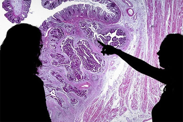
It is well known that early and accurate diagnosis of cancer is essential for both getting the correct treatment and obtaining the best outcome. At the first sign of trouble a biopsy is normally taken to examine tissue from suspicious lumps or lesions. A pathologist will then stain the tissue and examine it through a conventional microscope.
Pathology services, however, are increasingly under strain. The number of pathologists is decreasing year-on-year by approximately 15%. In addition, many cancers, such as prostate, are age related. Given that we have an aging population, there is an ever-increasing number of samples to be analysed.
Back in 2016, Cancer Research UK reported that ‘diagnostic services, including pathology, urgently need support and investment to ensure that diagnoses aren’t delayed and patients benefit from the latest treatment, and separately that ‘Immediate action is needed to avert a crisis in pathology capacity and ensure we have a service that is fit for the future’.
The government has suggested histopathology is ‘a key area ripe for technological revolution’. Part of that technological revolution is occurring in the form of Artificial Intelligence (AI). As we move from looking under a microscope to taking a digital image, pathologists are able to use AI to analyse these images and pick out key features that are indicative of cancer. These new analysis methods can help the pathologist in making the correct diagnosis.
AI, however, is not the only technology that is being explored. New spectroscopic microscopes are being developed that do not require any stains or dyes to “see” the tissue. The image is created by analysing vibrations in molecules that make up the tissue. AI can then be used to probe these chemical maps and look for features that cannot be seen under a conventional microscope.
These new techniques are very much in the developmental stage, but it is hoped that such methods will soon be available to help pathologists and improve cancer care.
Professor Peter Gardner FRSC, AMIChemE
Peter Gardner is a Professor of Analytical and Biomedical Spectroscopy in the Department of Chemical Engineering and the Photon Science Institute at the University of Manchester. He has previously held research positions at the Max Planck Institute in Berlin and the University of Cambridge.
Peter is a physical chemist with 40 years of research experience in the field of infrared and Raman spectroscopy. In 1999 he turned his attention to applying infrared microscopy to the analysis of cells and tissue. He has been working closely with the Christie hospital in Manchester and the Cancer Research UK Manchester Institute, developing new methods to analyse tissue samples from biopsies with a view to helping pathologists better diagnose cancer. He is leader of a UK network bringing together the communities of spectral pathology, digital pathology and computational pathology/AI.


